Flexible and Customizable
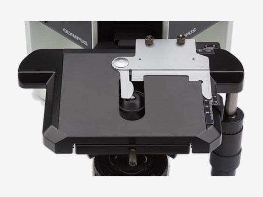
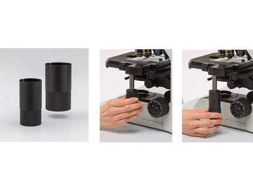
The LED illuminator for the BX53 is equivalent to or better than a 100 W halogen lamp, delivering brightness that’s appropriate for teaching or contrast methods. The BX53 microscope can be customized for different observation methods, such as phase contrast and fluorescence, with modular components.
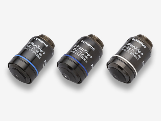
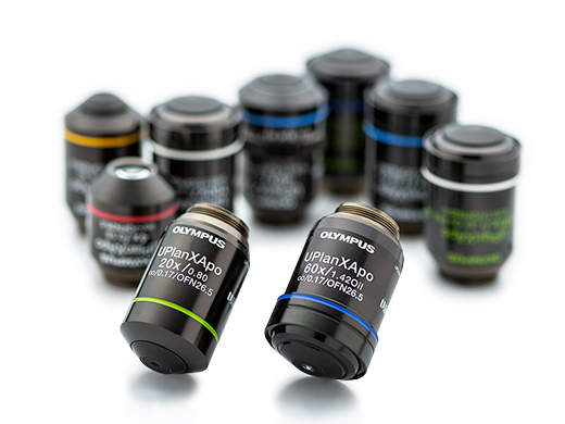
Acquire Precise Images with X Line ObjectivesThese objectives combine improved flatness, numerical aperture, and chromatic aberration to deliver clear, high-resolution images with better color accuracy across the entire spectrum. The elimination of violet color aberration creates clear whites and vivid pinks, improving contrast and sharpness. |
 |
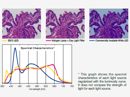 |
Bright LED Lighting Designed for Pathology and LaboratoryWith spectral characteristics similar to halogen light sources, the LED illuminator enables you to view the purple, cyan, and pink colors important in clinical research applications while enjoying the long use life of an LED. |
|---|
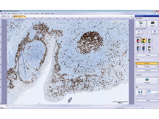
Digital Imaging Helps Simplify Your WorkFrom advanced research to capturing images for conferences, our digital microscope cameras and cellSens imaging software help ensure fluorescence imaging with a high signal-to-noise ratio. |
 |
|---|
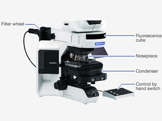 |
Upgrade to Motorized ComponentsAutomatically record and share magnification setting information with the optional coded nosepiece or change observation methods with a single touch using the hand switch. Motorized options include:
|
|---|
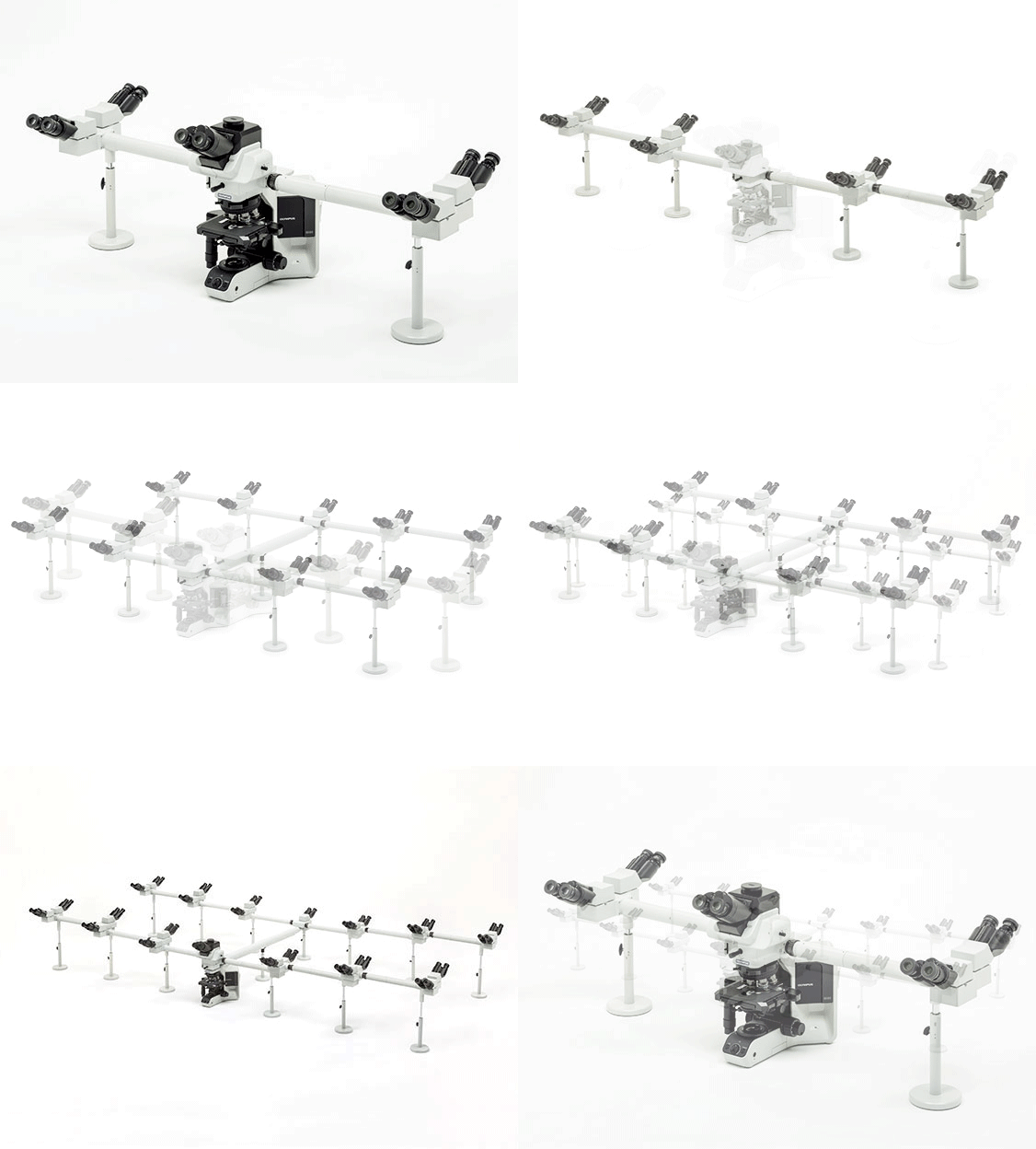
Bright Images in Multi-Head ConfigurationsThe multi-head light path has been redesigned not only to orient all images the same way but also to allow the intense LED to provide clear, bright images for up to 26 participants. |
|
|---|
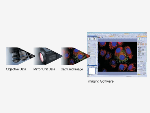 |
Save Microscopy Data via Coded UnitsAdd an optional coded nosepiece to your BX53 microscope to automatically record and share magnification setting information for post-imaging treatments. The metadata is automatically sent to cellSens software, helping minimize mistakes and scaling errors |
|---|


